Peer Review History
| Original SubmissionOctober 24, 2023 |
|---|
|
PONE-D-23-34916Vericiguat Suppresses Ventricular Tachyarrhythmias Inducibility in a Rabbit Myocardial Infarction ModelPLOS ONE Dear Dr. Chou, Thank you for submitting your manuscript to PLOS ONE. After careful consideration, we feel that it has merit but does not fully meet PLOS ONE’s publication criteria as it currently stands. Therefore, we invite you to submit a revised version of the manuscript that addresses the points raised during the review process. Please submit your revised manuscript by Feb 02 2024 11:59PM. If you will need more time than this to complete your revisions, please reply to this message or contact the journal office at plosone@plos.org. When you're ready to submit your revision, log on to https://www.editorialmanager.com/pone/ and select the 'Submissions Needing Revision' folder to locate your manuscript file. Please include the following items when submitting your revised manuscript:
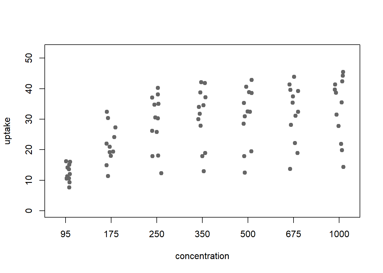
If you would like to make changes to your financial disclosure, please include your updated statement in your cover letter. Guidelines for resubmitting your figure files are available below the reviewer comments at the end of this letter. If applicable, we recommend that you deposit your laboratory protocols in protocols.io to enhance the reproducibility of your results. Protocols.io assigns your protocol its own identifier (DOI) so that it can be cited independently in the future. For instructions see: https://journals.plos.org/plosone/s/submission-guidelines#loc-laboratory-protocols. Additionally, PLOS ONE offers an option for publishing peer-reviewed Lab Protocol articles, which describe protocols hosted on protocols.io. Read more information on sharing protocols at https://plos.org/protocols?utm_medium=editorial-email&utm_source=authorletters&utm_campaign=protocols. We look forward to receiving your revised manuscript. Kind regards, Elena G. Tolkacheva, PhD Academic Editor PLOS ONE Journal Requirements: When submitting your revision, we need you to address these additional requirements. 1. Please ensure that your manuscript meets PLOS ONE's style requirements, including those for file naming. The PLOS ONE style templates can be found at https://journals.plos.org/plosone/s/file?id=wjVg/PLOSOne_formatting_sample_main_body.pdf and 2. To comply with PLOS ONE submissions requirements, in your Methods section, please provide additional information regarding the experiments involving animals and ensure you have included details on (1) methods of sacrifice and (2) efforts to alleviate suffering. 3. We note that the grant information you provided in the ‘Funding Information’ and ‘Financial Disclosure’ sections do not match. When you resubmit, please ensure that you provide the correct grant numbers for the awards you received for your study in the ‘Funding Information’ section. 4. Thank you for stating the following financial disclosure: "This study was supported by the Ministry of Science and Technology, Taiwan (110-2314-B-182A-119- to P.C. Chang) and Chang Gung Medical Foundation (CMRPG3L1202 to P.C. Chang)." Please state what role the funders took in the study. If the funders had no role, please state: ""The funders had no role in study design, data collection and analysis, decision to publish, or preparation of the manuscript."" If this statement is not correct you must amend it as needed. Please include this amended Role of Funder statement in your cover letter; we will change the online submission form on your behalf. 5. Please include captions for your Supporting Information files at the end of your manuscript, and update any in-text citations to match accordingly. Please see our Supporting Information guidelines for more information: http://journals.plos.org/plosone/s/supporting-information. Additional Editor Comments: Please address comments indicated by the Reviewers. [Note: HTML markup is below. Please do not edit.] Reviewers' comments: Reviewer's Responses to Questions Comments to the Author 1. Is the manuscript technically sound, and do the data support the conclusions? The manuscript must describe a technically sound piece of scientific research with data that supports the conclusions. Experiments must have been conducted rigorously, with appropriate controls, replication, and sample sizes. The conclusions must be drawn appropriately based on the data presented. Reviewer #1: Partly Reviewer #2: Yes ********** 2. Has the statistical analysis been performed appropriately and rigorously? Reviewer #1: Yes Reviewer #2: Yes ********** 3. Have the authors made all data underlying the findings in their manuscript fully available? The PLOS Data policy requires authors to make all data underlying the findings described in their manuscript fully available without restriction, with rare exception (please refer to the Data Availability Statement in the manuscript PDF file). The data should be provided as part of the manuscript or its supporting information, or deposited to a public repository. For example, in addition to summary statistics, the data points behind means, medians and variance measures should be available. If there are restrictions on publicly sharing data—e.g. participant privacy or use of data from a third party—those must be specified. Reviewer #1: Yes Reviewer #2: No ********** 4. Is the manuscript presented in an intelligible fashion and written in standard English? PLOS ONE does not copyedit accepted manuscripts, so the language in submitted articles must be clear, correct, and unambiguous. Any typographical or grammatical errors should be corrected at revision, so please note any specific errors here. Reviewer #1: Yes Reviewer #2: Yes ********** 5. Review Comments to the Author Please use the space provided to explain your answers to the questions above. You may also include additional comments for the author, including concerns about dual publication, research ethics, or publication ethics. (Please upload your review as an attachment if it exceeds 20,000 characters) Reviewer #1: The present study by Chang et al. utilises the optical mapping technique to investigate how the drug vericiguat, which has recently been approved for the use in HFrEF, affects electrical activity and calcium handling in a rabbit model of myocardial infarction. The authors show that this compound reduced the number of premature ventricular beats in this model, as well as causing alterations in conduction velocity, action potential characteristics, and calcium transients. In general, the experiments have been carried out well, however i do have a number of comments/questions. 1) I am a little confused regarding the animals which underwent the repeated EP study, and then underwent the drug challenge. These are your time controls- so this is great that these have been done- but then are these hearts when drug-perfused also included in the analysis? If so this would mean they would have been on the optical mapping set up even longer than what is shown as the time control (as it would have been control-control-drug, instead of control-drug). Is it these animals that were driving the significance? This needs to be described better in the text, and n numbers should be included in the text and figure legends when describing these. 2) Did the Authors attempt to wash out vericiguat on these preperations? 3) I find it a shame that there are no sham operated animals, as it would have been nice to compare the effects in MI vs Sham. Have these experiments been carried out? If so they need to be included, otherwise there needs to be an addition to the limitations that these experiments were not done. 4) On the mapping isochrones, you clearly label the MI region. Was this all scar tissue, and thats how you defined this? Did you ever quantify the area affected by the MI? Was this similar with all animals? This also brings me onto the methods, where it is stated that either one or two branches of the LCA were ligated. What was the determining factor here, if one or two branches were ligated? 5) When the hearts were being optically imaged, was the flow rate controlled? Did the Authors notice any difference in the amount of liquid coming out- or was this measured- this could be interesting to know, to see if any effects are caused by alterations in vessel diameter etc, as opposed to being due to direct effects on the myocardium. 6) How long was the drug perfused for? It states the time controls were 20 minutes, so did you record the drug records after 20 mins of perfusion. Again, this needs to be clarified in the manuscript. 7) The differences seen in CV, for example, are very small, despite there being significance. Would these small differences actually be physiologically relevant? I think there needs to be some discussion of this 8) In all bar charts, i would like to see the individual data points (perhaps even joining up the before and after drug numbers, to see exactly the trends on an individual heart level) 9) Was Echo carried out pre-MI? It would be nice to see these data too- just to show that there is a decrease in EF. 10) n numbers should be included in all the figure legends 11) When examples are shown in figures comparing pre and post drug, are these taken from the same animal. This should be emphasised in the figure legend as well) 12) Please check the units in some of the figures. For example, in Figure 3, the bar chart states that CV is in cm/ms when i believe it should be in cm/s as it is on the isochrones.. 13) I believe the figure legend on Supplementary Figure 2 is incorrect, as I believe this is showing the time control data, but in the legend it mentions the drug treatment. 14) Please check for repetition in the manuscript. For example on lines 209-212, there is discussion again about how the model was made, which maybe isnt really needed at this point. 15) Please check grammar throughout the manuscript. For example in the results section of the abstract it states 'accelerated of intracellular calcium' when it should be 'acceleration of intracellular calcium'. Also there should be 'duration' after action potential in this section. 16) I also think there needs to be a number of additional references added- for example on line 59, when stating the fact that vericiguat has been proven to be beneficial in HF patients. 17) I also wondered why the Authors utilised Labview for analysis, when there are a number of free analysis programs available for analysing these data - for example ElectroMap, Rhythm or COSMAS. 18) When discussing the potential mechanisms of the alterations in Calcium transient, i believe there also needs to be a mention of NCX. Although this is discussed later on, when discussing the tau of the transient, this is one of the major players here. Also, im not sure the L-type Ca current should be mentioned here, as this will be having minimal effects on the value of the downstroke of the transient. 19) Where the rabbits male or female or a mix- this needs to be described in the methods. 20) Im guessing the blebbistatin was included in the Tyrodes solution for the imaging part, and this was continuous? This should be a bit clearer in the text i believe. 21) When the affects of NO are discussed on the sodium current, is this the peak sodium current, or the late component. I believe there needs to be some discussion of that, as it may have implications for NCX, especially if its the late component.. 22) At the end of the discussion of IKr vs INa, I believe there should be there needs to be an acknowledgement that other ion channels may also play a role (line 279) 23) When discussing ICa, the authors often discuss beta-adrenergic stimulation. As the Authors did not use this method of inducing arrrhythmias in this model, im not really sure these data can be used as an argument? Did you ever try these experiments in the presence of Isoproterenol for example? 24) I know you show the tape measure in Figure 5A, but please put a scale bar on. Also it may be nice to show this without the MI portion faded out, so the reader can see what this looks like- or at least put another photo of this heart without the MI portion greyed out. Reviewer #2: In their study “Vericiguat suppresses ventricular tachyarrhythmias inducibility in a rabbit myocardial infarction model” Chou et al. investigate the effect of a vericiguat, a stimulator of guanylate cyclase, onto the susceptibility of infarcted isolated rabbit hearts to ventricular arrhythmias using optical mapping. The study is examplary for demonstrating the utility of optical mapping for studying the mechanisms underlying the arrhythmogenesis of compounds and disease in electrophysiological studies. The study is scientifically sound, rigorously executed and includes a detailed discussion putting their findings into perspective. I have only minor concerns regarding some of the methodological approaches and presentation of the results: - Please use a format with figures and figure captions in place with figure captions next to the figure and the figure close to where referenced in the text. That way I do not have to scroll back and forth from the beginning to the end of the document all the time. - The study lacks a conflict of interest statement. - Please add age and sex of rabbits. - Are there representative APD maps that complement the bar plot in Fig. 2A? - Please add the number of hearts in Fig. 2 A-E. - The bar plots do not reveal the number of individual measurements and their spread. Please use a scatter or stripchart plot in addition to bar plots, e.g. as shown here: https://www.ashander.info/notes/barchart-alternatives-in-base-R_files/figure-html/scatter-v-factors-1.png - Please add an overlay of the traces for one AP or CA-transient so that one can see the effect more clearly or align them in time. The traces as they are presented right now are not helpful for evaluating a change in duration. - line 186: I feel like the statement “vericiguat therapy significantly slowed the ventricular myocardial conduction” is not supported by the presented data. In the bar plot in Fig. 3A,B the difference is hardly noticeable and I am surprised it is significant based on the p-value (which is a poor measure for statistical significance) given the large error bars. See 2 points below. - Fig. 3. I do not understand how CV was measured. Please add points from where to where and along which path the distance and CV was measured in the figure. - Given that the change in CV in the bar plots is not very large: Could this measurement be improved? For instance, the authors could measure the distance between isochrones for every point on the isochrone and compute a CV map and average over the data. Maybe this would produce a more distinct separation between baseline and Vericiquat. - Fig. 3: Why are there Ca alternates but no V alternates? - lines 195-198 and Fig. 4B: I would like to see more representative traces that highlight the shortening of the tau value with overlays of the baseline and vericiguat traces. - Please provide videos for optical mapping data. - Please provide the video raw data in an appropriate format (e.g. a Matlab or Numpy .mat / .npy file which includes an array) for Fig. 5 D, G. - line 233: how did the shortening of tau / acceleration of Ca homeostasis contribute to suppression of alternans? - line 58: missing reference - line 48: coronary artery disease (CAD) contributes - lines 318: Why did the authors choose this much higher concentration and what happens when the dosage is reduced, let’s say to 1 micro mol/L? - line 323: missing reference - In lines 298-299 the authors mention electrophysiological changes in ischemic failing hearts including slowing of CV and prolongation of AP, which they also report with vericiguat. How can these changes be pro- and anti-arrhythmic at the same time? ********** 6. PLOS authors have the option to publish the peer review history of their article (what does this mean?). If published, this will include your full peer review and any attached files. If you choose “no”, your identity will remain anonymous but your review may still be made public. Do you want your identity to be public for this peer review? For information about this choice, including consent withdrawal, please see our Privacy Policy. Reviewer #1: No Reviewer #2: No ********** [NOTE: If reviewer comments were submitted as an attachment file, they will be attached to this email and accessible via the submission site. Please log into your account, locate the manuscript record, and check for the action link "View Attachments". If this link does not appear, there are no attachment files.] While revising your submission, please upload your figure files to the Preflight Analysis and Conversion Engine (PACE) digital diagnostic tool, https://pacev2.apexcovantage.com/. PACE helps ensure that figures meet PLOS requirements. To use PACE, you must first register as a user. Registration is free. Then, login and navigate to the UPLOAD tab, where you will find detailed instructions on how to use the tool. If you encounter any issues or have any questions when using PACE, please email PLOS at figures@plos.org. Please note that Supporting Information files do not need this step. |
| Revision 1 |
|
PONE-D-23-34916R1Vericiguat Suppresses Ventricular Tachyarrhythmias Inducibility in a Rabbit Myocardial Infarction ModelPLOS ONE Dear Dr. Chou, Thank you for submitting your manuscript to PLOS ONE. After careful consideration, we feel that it has merit but does not fully meet PLOS ONE’s publication criteria as it currently stands. Therefore, we invite you to submit a revised version of the manuscript that addresses the points raised during the review process. Please submit your revised manuscript by Mar 30 2024 11:59PM. If you will need more time than this to complete your revisions, please reply to this message or contact the journal office at plosone@plos.org. When you're ready to submit your revision, log on to https://www.editorialmanager.com/pone/ and select the 'Submissions Needing Revision' folder to locate your manuscript file. Please include the following items when submitting your revised manuscript:
If applicable, we recommend that you deposit your laboratory protocols in protocols.io to enhance the reproducibility of your results. Protocols.io assigns your protocol its own identifier (DOI) so that it can be cited independently in the future. For instructions see: https://journals.plos.org/plosone/s/submission-guidelines#loc-laboratory-protocols. Additionally, PLOS ONE offers an option for publishing peer-reviewed Lab Protocol articles, which describe protocols hosted on protocols.io. Read more information on sharing protocols at https://plos.org/protocols?utm_medium=editorial-email&utm_source=authorletters&utm_campaign=protocols. We look forward to receiving your revised manuscript. Kind regards, Elena G. Tolkacheva, PhD Academic Editor PLOS ONE Journal Requirements: Please review your reference list to ensure that it is complete and correct. If you have cited papers that have been retracted, please include the rationale for doing so in the manuscript text, or remove these references and replace them with relevant current references. Any changes to the reference list should be mentioned in the rebuttal letter that accompanies your revised manuscript. If you need to cite a retracted article, indicate the article’s retracted status in the References list and also include a citation and full reference for the retraction notice. Additional Editor Comments: Please address minor comments indicated by the reviewer. [Note: HTML markup is below. Please do not edit.] Reviewers' comments: Reviewer's Responses to Questions Comments to the Author 1. If the authors have adequately addressed your comments raised in a previous round of review and you feel that this manuscript is now acceptable for publication, you may indicate that here to bypass the “Comments to the Author” section, enter your conflict of interest statement in the “Confidential to Editor” section, and submit your "Accept" recommendation. Reviewer #1: All comments have been addressed Reviewer #2: (No Response) ********** 2. Is the manuscript technically sound, and do the data support the conclusions? The manuscript must describe a technically sound piece of scientific research with data that supports the conclusions. Experiments must have been conducted rigorously, with appropriate controls, replication, and sample sizes. The conclusions must be drawn appropriately based on the data presented. Reviewer #1: Yes Reviewer #2: Yes ********** 3. Has the statistical analysis been performed appropriately and rigorously? Reviewer #1: Yes Reviewer #2: I Don't Know ********** 4. Have the authors made all data underlying the findings in their manuscript fully available? The PLOS Data policy requires authors to make all data underlying the findings described in their manuscript fully available without restriction, with rare exception (please refer to the Data Availability Statement in the manuscript PDF file). The data should be provided as part of the manuscript or its supporting information, or deposited to a public repository. For example, in addition to summary statistics, the data points behind means, medians and variance measures should be available. If there are restrictions on publicly sharing data—e.g. participant privacy or use of data from a third party—those must be specified. Reviewer #1: Yes Reviewer #2: No ********** 5. Is the manuscript presented in an intelligible fashion and written in standard English? PLOS ONE does not copyedit accepted manuscripts, so the language in submitted articles must be clear, correct, and unambiguous. Any typographical or grammatical errors should be corrected at revision, so please note any specific errors here. Reviewer #1: Yes Reviewer #2: Yes ********** 6. Review Comments to the Author Please use the space provided to explain your answers to the questions above. You may also include additional comments for the author, including concerns about dual publication, research ethics, or publication ethics. (Please upload your review as an attachment if it exceeds 20,000 characters) Reviewer #1: All comments have been addressed which i thank the Authors for. Please check grammar and spelling throughout the manuscript though- for instance on line 272, cGC is stated instead of sGC.. Reviewer #2: Thank you very much for the detailed response and revision of the manuscript. I have a few remaining minor concerns, the main concern being that the measured effects (APD / CV) are very small and I would like to verify the effect in the raw data myself (PlosONE specifically asks me whether all raw data was made available). - Please mark all revised parts of your manuscript in blue, otherwise it is difficult to confirm which parts were revised. - Thank you very much for providing raw data (Pacing induced VT raw data for Fig 5). Could you please provide raw data for Figs. 2 and 3 which provides a comparison of baseline and Vericiguat? Please provide several paired corresponding videos with baseline and Vericiguat so that I can reproduce and verify the plots in Figs. 2 and 3. - The authors should phrase the significance of the measured APD and CV changes more carefully. They may be statistically significant, but still small. - The rabbits are very young (6 months). The limitations section should list this as a potential limitation (would the results change with older rabbits?). As a note: You can use the following software for the post-processing of optical mapping videos: https://github.com/cardiacvision/optimap Tutorial 13 explains how to read in .rsh videos: https://optimap.readthedocs.io/en/latest/tutorials/io/ ********** 7. PLOS authors have the option to publish the peer review history of their article (what does this mean?). If published, this will include your full peer review and any attached files. If you choose “no”, your identity will remain anonymous but your review may still be made public. Do you want your identity to be public for this peer review? For information about this choice, including consent withdrawal, please see our Privacy Policy. Reviewer #1: No Reviewer #2: No ********** [NOTE: If reviewer comments were submitted as an attachment file, they will be attached to this email and accessible via the submission site. Please log into your account, locate the manuscript record, and check for the action link "View Attachments". If this link does not appear, there are no attachment files.] While revising your submission, please upload your figure files to the Preflight Analysis and Conversion Engine (PACE) digital diagnostic tool, https://pacev2.apexcovantage.com/. PACE helps ensure that figures meet PLOS requirements. To use PACE, you must first register as a user. Registration is free. Then, login and navigate to the UPLOAD tab, where you will find detailed instructions on how to use the tool. If you encounter any issues or have any questions when using PACE, please email PLOS at figures@plos.org. Please note that Supporting Information files do not need this step. |
| Revision 2 |
|
PONE-D-23-34916R2Vericiguat Suppresses Ventricular Tachyarrhythmias Inducibility in a Rabbit Myocardial Infarction ModelPLOS ONE Dear Dr. Chou, Thank you for submitting your manuscript to PLOS ONE. After careful consideration, we feel that it has merit but does not fully meet PLOS ONE’s publication criteria as it currently stands. Therefore, we invite you to submit a revised version of the manuscript that addresses the points raised during the review process. Please address comments indicated by the Reviewer. Please submit your revised manuscript by Apr 26 2024 11:59PM. If you will need more time than this to complete your revisions, please reply to this message or contact the journal office at plosone@plos.org. When you're ready to submit your revision, log on to https://www.editorialmanager.com/pone/ and select the 'Submissions Needing Revision' folder to locate your manuscript file. Please include the following items when submitting your revised manuscript:
If applicable, we recommend that you deposit your laboratory protocols in protocols.io to enhance the reproducibility of your results. Protocols.io assigns your protocol its own identifier (DOI) so that it can be cited independently in the future. For instructions see: https://journals.plos.org/plosone/s/submission-guidelines#loc-laboratory-protocols. Additionally, PLOS ONE offers an option for publishing peer-reviewed Lab Protocol articles, which describe protocols hosted on protocols.io. Read more information on sharing protocols at https://plos.org/protocols?utm_medium=editorial-email&utm_source=authorletters&utm_campaign=protocols. We look forward to receiving your revised manuscript. Kind regards, Elena G. Tolkacheva, PhD Academic Editor PLOS ONE Journal Requirements: Please review your reference list to ensure that it is complete and correct. If you have cited papers that have been retracted, please include the rationale for doing so in the manuscript text, or remove these references and replace them with relevant current references. Any changes to the reference list should be mentioned in the rebuttal letter that accompanies your revised manuscript. If you need to cite a retracted article, indicate the article’s retracted status in the References list and also include a citation and full reference for the retraction notice. [Note: HTML markup is below. Please do not edit.] Reviewers' comments: Reviewer's Responses to Questions Comments to the Author 1. If the authors have adequately addressed your comments raised in a previous round of review and you feel that this manuscript is now acceptable for publication, you may indicate that here to bypass the “Comments to the Author” section, enter your conflict of interest statement in the “Confidential to Editor” section, and submit your "Accept" recommendation. Reviewer #2: (No Response) ********** 2. Is the manuscript technically sound, and do the data support the conclusions? The manuscript must describe a technically sound piece of scientific research with data that supports the conclusions. Experiments must have been conducted rigorously, with appropriate controls, replication, and sample sizes. The conclusions must be drawn appropriately based on the data presented. Reviewer #2: Partly ********** 3. Has the statistical analysis been performed appropriately and rigorously? Reviewer #2: No ********** 4. Have the authors made all data underlying the findings in their manuscript fully available? The PLOS Data policy requires authors to make all data underlying the findings described in their manuscript fully available without restriction, with rare exception (please refer to the Data Availability Statement in the manuscript PDF file). The data should be provided as part of the manuscript or its supporting information, or deposited to a public repository. For example, in addition to summary statistics, the data points behind means, medians and variance measures should be available. If there are restrictions on publicly sharing data—e.g. participant privacy or use of data from a third party—those must be specified. Reviewer #2: No ********** 5. Is the manuscript presented in an intelligible fashion and written in standard English? PLOS ONE does not copyedit accepted manuscripts, so the language in submitted articles must be clear, correct, and unambiguous. Any typographical or grammatical errors should be corrected at revision, so please note any specific errors here. Reviewer #2: Yes ********** 6. Review Comments to the Author Please use the space provided to explain your answers to the questions above. You may also include additional comments for the author, including concerns about dual publication, research ethics, or publication ethics. (Please upload your review as an attachment if it exceeds 20,000 characters) Reviewer #2: Thank you very much for providing the requested raw data. I confirm that the APD prolongation occurs with Vericiguat in all data I reviewed. However, I cannot confirm the findings regarding the change in CV. I attached 2 videos showing a comparison of the wave propagation at Baseline vs. Vericiguat (each in rabbits 1 and 2, recordings 200.rsh). In rabbit 1, I do not notice any difference in the wave speed and in rabbit 2 the wave activates the ventricle with Vericiguat first, even though the wave comes in later, suggesting transmural conduction or a much faster CV with Vericiguat. In any way, the data I reviewed does not support that Vericiguat decreases CV, and I therefore suggest removing the data and claim or revising the paper and presenting the analysis in a convincing fashion. I leave this to the authors and recommend' accept' if the CV data is removed. ********** 7. PLOS authors have the option to publish the peer review history of their article (what does this mean?). If published, this will include your full peer review and any attached files. If you choose “no”, your identity will remain anonymous but your review may still be made public. Do you want your identity to be public for this peer review? For information about this choice, including consent withdrawal, please see our Privacy Policy. Reviewer #2: No ********** [NOTE: If reviewer comments were submitted as an attachment file, they will be attached to this email and accessible via the submission site. Please log into your account, locate the manuscript record, and check for the action link "View Attachments". If this link does not appear, there are no attachment files.] While revising your submission, please upload your figure files to the Preflight Analysis and Conversion Engine (PACE) digital diagnostic tool, https://pacev2.apexcovantage.com/. PACE helps ensure that figures meet PLOS requirements. To use PACE, you must first register as a user. Registration is free. Then, login and navigate to the UPLOAD tab, where you will find detailed instructions on how to use the tool. If you encounter any issues or have any questions when using PACE, please email PLOS at figures@plos.org. Please note that Supporting Information files do not need this step. |
| Revision 3 |
|
Vericiguat Suppresses Ventricular Tachyarrhythmias Inducibility in a Rabbit Myocardial Infarction Model PONE-D-23-34916R3 Dear Dr. Chou, We’re pleased to inform you that your manuscript has been judged scientifically suitable for publication and will be formally accepted for publication once it meets all outstanding technical requirements. Within one week, you’ll receive an e-mail detailing the required amendments. When these have been addressed, you’ll receive a formal acceptance letter and your manuscript will be scheduled for publication. An invoice will be generated when your article is formally accepted. Please note, if your institution has a publishing partnership with PLOS and your article meets the relevant criteria, all or part of your publication costs will be covered. Please make sure your user information is up-to-date by logging into Editorial Manager at Editorial Manager® and clicking the ‘Update My Information' link at the top of the page. If you have any questions relating to publication charges, please contact our Author Billing department directly at authorbilling@plos.org. If your institution or institutions have a press office, please notify them about your upcoming paper to help maximize its impact. If they’ll be preparing press materials, please inform our press team as soon as possible -- no later than 48 hours after receiving the formal acceptance. Your manuscript will remain under strict press embargo until 2 pm Eastern Time on the date of publication. For more information, please contact onepress@plos.org. Kind regards, Elena G. Tolkacheva, PhD Academic Editor PLOS ONE Additional Editor Comments (optional): Reviewers' comments: Reviewer's Responses to Questions Comments to the Author 1. If the authors have adequately addressed your comments raised in a previous round of review and you feel that this manuscript is now acceptable for publication, you may indicate that here to bypass the “Comments to the Author” section, enter your conflict of interest statement in the “Confidential to Editor” section, and submit your "Accept" recommendation. Reviewer #2: All comments have been addressed ********** 2. Is the manuscript technically sound, and do the data support the conclusions? The manuscript must describe a technically sound piece of scientific research with data that supports the conclusions. Experiments must have been conducted rigorously, with appropriate controls, replication, and sample sizes. The conclusions must be drawn appropriately based on the data presented. Reviewer #2: Yes ********** 3. Has the statistical analysis been performed appropriately and rigorously? Reviewer #2: I Don't Know ********** 4. Have the authors made all data underlying the findings in their manuscript fully available? The PLOS Data policy requires authors to make all data underlying the findings described in their manuscript fully available without restriction, with rare exception (please refer to the Data Availability Statement in the manuscript PDF file). The data should be provided as part of the manuscript or its supporting information, or deposited to a public repository. For example, in addition to summary statistics, the data points behind means, medians and variance measures should be available. If there are restrictions on publicly sharing data—e.g. participant privacy or use of data from a third party—those must be specified. Reviewer #2: No ********** 5. Is the manuscript presented in an intelligible fashion and written in standard English? PLOS ONE does not copyedit accepted manuscripts, so the language in submitted articles must be clear, correct, and unambiguous. Any typographical or grammatical errors should be corrected at revision, so please note any specific errors here. Reviewer #2: Yes ********** 6. Review Comments to the Author Please use the space provided to explain your answers to the questions above. You may also include additional comments for the author, including concerns about dual publication, research ethics, or publication ethics. (Please upload your review as an attachment if it exceeds 20,000 characters) Reviewer #2: Thank you very much for removing the part about CV. Please check the PLOS data policy and upload the supporting raw data to a repository before publication. ********** 7. PLOS authors have the option to publish the peer review history of their article (what does this mean?). If published, this will include your full peer review and any attached files. If you choose “no”, your identity will remain anonymous but your review may still be made public. Do you want your identity to be public for this peer review? For information about this choice, including consent withdrawal, please see our Privacy Policy. Reviewer #2: No ********** |
| Formally Accepted |
|
PONE-D-23-34916R3 PLOS ONE Dear Dr. Chou, I'm pleased to inform you that your manuscript has been deemed suitable for publication in PLOS ONE. Congratulations! Your manuscript is now being handed over to our production team. At this stage, our production department will prepare your paper for publication. This includes ensuring the following: * All references, tables, and figures are properly cited * All relevant supporting information is included in the manuscript submission, * There are no issues that prevent the paper from being properly typeset If revisions are needed, the production department will contact you directly to resolve them. If no revisions are needed, you will receive an email when the publication date has been set. At this time, we do not offer pre-publication proofs to authors during production of the accepted work. Please keep in mind that we are working through a large volume of accepted articles, so please give us a few weeks to review your paper and let you know the next and final steps. Lastly, if your institution or institutions have a press office, please let them know about your upcoming paper now to help maximize its impact. If they'll be preparing press materials, please inform our press team within the next 48 hours. Your manuscript will remain under strict press embargo until 2 pm Eastern Time on the date of publication. For more information, please contact onepress@plos.org. If we can help with anything else, please email us at customercare@plos.org. Thank you for submitting your work to PLOS ONE and supporting open access. Kind regards, PLOS ONE Editorial Office Staff on behalf of Dr. Elena G. Tolkacheva Academic Editor PLOS ONE |
Open letter on the publication of peer review reports
PLOS recognizes the benefits of transparency in the peer review process. Therefore, we enable the publication of all of the content of peer review and author responses alongside final, published articles. Reviewers remain anonymous, unless they choose to reveal their names.
We encourage other journals to join us in this initiative. We hope that our action inspires the community, including researchers, research funders, and research institutions, to recognize the benefits of published peer review reports for all parts of the research system.
Learn more at ASAPbio .